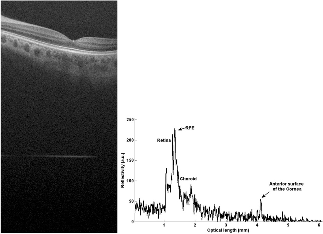
Fig. 2.
Composite A-scan of low myopic human eye in vivo. The retina is located close to the origin of the graph and the front surface of the cornea further away. (a) gives the cross section OCT image, which contains the retinal image and corneal signal. (b) is the A-line information for the IOL measurement. For this subject, the AL calculated by the formula is (35 – 2.236)/1.3375 = 24.496 mm. For comparison, length measurement data of myopic eyes with IOL master also has been conducted. The comparisons between our SD-OCT system and IOL master results are given in Table 1.

