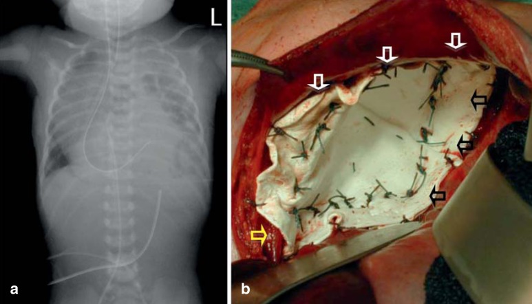
Figure 1.
Radiological and intraoperative appearance of left-sided congenital diaphragmatic hernia.
a) Postnatal chest radiograph of a neonate with left-sided congenital diaphragmatic hernia. Note the left-sided enterothorax due to the defect in the diaphragm, with a barely visible left lung (hypoplasia), and the intrathoracic location of the gastric tube.
b) Intraoperative appearance after closure of a large, overlapping conical GoreTex patch. The conical shape has decreased the thoracic dead space and increased the abdominal space, reducing the risk of abdominal compartment syndrome. At the anterior margin the ventral diaphragmatic rim can be seen (white arrows, and medially the left crus of the diaphragm is visible attached to the esophagus (yellow arrow). Dorsally, the patch is attached around the ribs (black arrows)

