Figure 2.
TSN1 and TSN2 Localize to SGs and PBs.
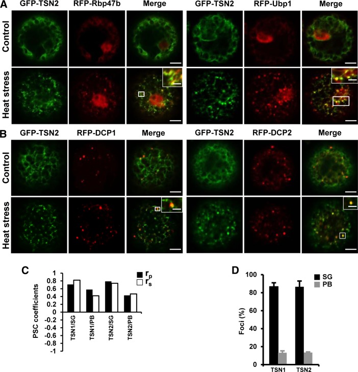
(A) and (B) Colocalization of GFP-TSN2 (green) with the SG (A) and PB (B) marker proteins (red) RFP-Rbp47b/RFP-Ubp1 and RFP-DCP1/RFP-DCP2, respectively, in protoplasts from N. benthamiana under control conditions (23°C) or after 30 min at 39°C (heat stress). N-terminal GFP and RFP fusion proteins were expressed under the control of the 35S promoter. White boxes indicate the areas that are magnified in the insets (merge). For colocalization of GFP-TSN1 with the SG and PB markers, see Supplemental Figure 1. Bars = 5 μm (2 μm in insets).
(C) Pearson and Spearman coefficients (rp and rs, respectively) of colocalization (PSC; French et al., 2008) of GFP-TSN1 and GFP-TSN2 with the SG marker RFP-Rbp47b and the PB marker RFP-DCP1 in protoplasts after heat stress.
(D) Frequency (%) of colocalization of TSN-containing foci with the SG marker RFP-Rbp47b and the PB marker RFP-DCP1 in protoplasts after heat stress. Data show means ± sd of triplicate experiments, each including 20 protoplasts.

