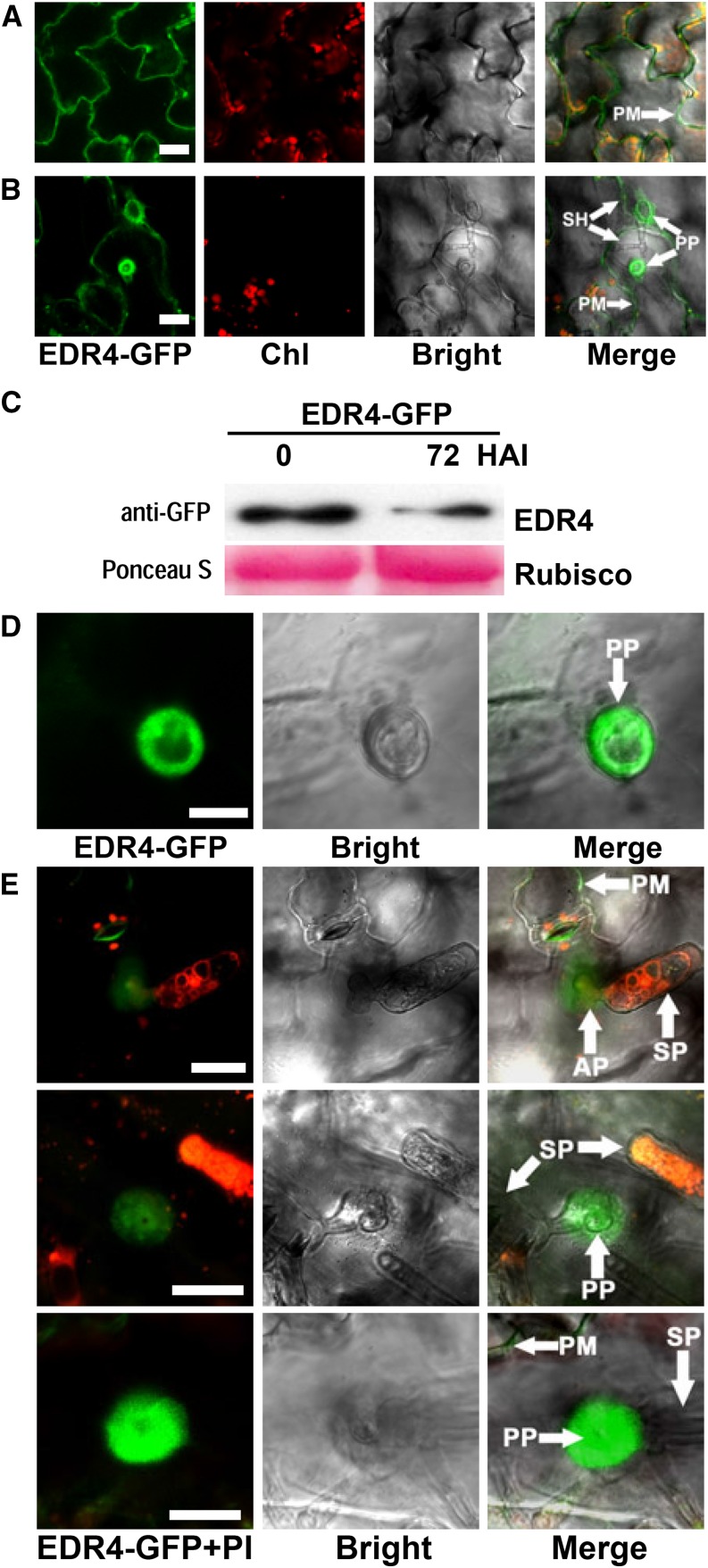
Figure 4.
EDR4-GFP Accumulates at the Powdery Mildew Penetration Site in Epidermal Cells.
(A) EDR4-GFP localization in epidermal cells of uninfected leaves. Bar = 25 μm.
(B) EDR4-GFP accumulates under the penetration peg of the secondary hyphal infection site in epidermal cells at 48 HAI with G. cichoracearum. Bar = 25 μm.
(C) Four-week-old transgenic EDR4-GFP plants were infected with G. cichoracearum. Immunoblotting was performed using an anti-GFP antibody. Ponceau S staining of Rubisco is shown as a loading control. The experiment was repeated at least three times with similar results.
(D) and (E) Accumulation of EDR4-GFP around the penetration peg of the appressorium at 24 HAI with G. cichoracearum, shown in different views. Leaves in (E) were stained with propidium iodide (PI) to visualize the fungal structures, which appear red. Bar in (D) = 10 μm; bars in (E) = 20 μm.
AP, appressoria; Chl, chloroplast; PM, plasma membrane; PP, penetration peg; SH, secondary hyphae; SP, spores.

