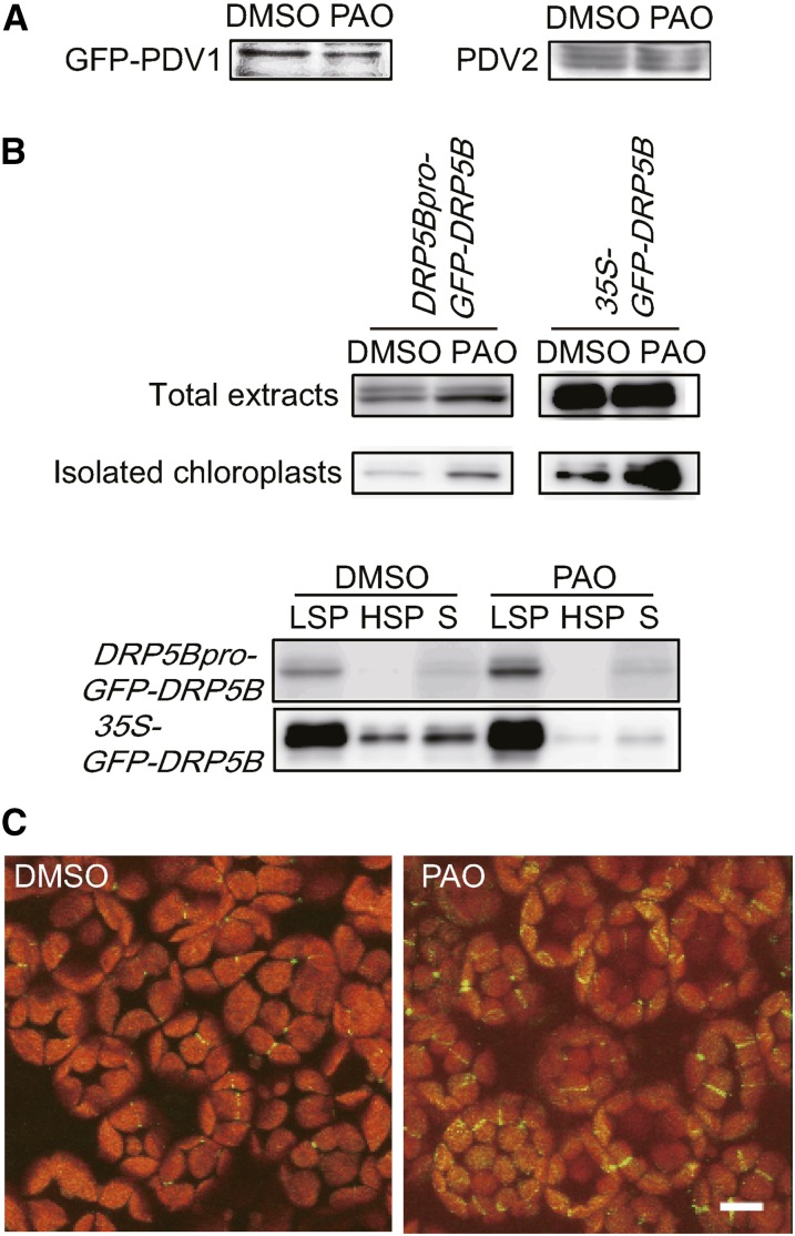
Figure 5.
PAO Treatment Increases DRP5B Expression and Enhances DRP5B Recruitment to Chloroplasts.
(A) Levels of GFP-PDV1 and PDV2 in pdv1 plants expressing GFP-PDV1 under the control of the PDV1 promoter. Plants were treated with DMSO or 25 μM PAO for 3 d. Total extracts containing 50 μg of protein were loaded for analysis of GFP-PDV1 and PDV2. GFP-PDV1 and PDV2 were detected with anti-GFP and anti-PDV2 antibodies, respectively.
(B) Levels of GFP-DRP5B in drp5b plants expressing GFP-DRP5B under the control of the DRP5B promoter (DRP5Bpro-GFP-DRP5B) and the cauliflower mosaic virus 35S promoter (35S-GFP-DRP5B). In total extracts, 50 and 2.5 μg of proteins extracted from whole plants were loaded for analysis of DRP5Bpro-GFP-DRP5B and 35S-GFP-DRP5B, respectively. In isolated chloroplasts, 2 and 0.2 μg of proteins were loaded for analysis of DRP5Bpro-GFP-DRP5B and 35S-GFP-DRP5B, respectively. GFP-DRP5B was detected with an anti-GFP antibody. In fractionated samples, homogenates prepared from 30 to 50 mg of leaves were fractionated into LSP, high-speed pellet (HSP), and supernatant (S) fractions by centrifugation. Two biological replicates showed equivalent results.
(C) DRP5Bpro-GFP-DRP5B drp5b plants were treated with DMSO (left panel) or 25 μM PAO (right panel) for 3 d. Images of GFP and chlorophyll fluorescence were taken using a confocal laser-scanning microscope. Bar = 10 μm.

