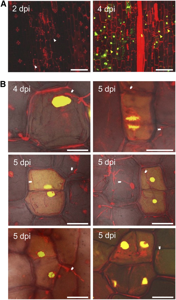
Figure 3.
U. maydis Induces DNA Synthesis in Infected Maize Seedlings.
(A) Maize seedlings were infected by U. maydis wild-type strain SG200, and then tissue was incubated in EdU to visualize in vivo DNA synthesis in the host cells. Samples were imaged at 2 and 4 DPI by confocal microscopy. Left, at 2 DPI, the fungal proliferation was observed subepidermally; host cells adjacent to fungal hyphae were considered to be colonized cells (white arrowheads). No EdU incorporation was observed. Right, at 4 DPI, numerous colonized cells showed EdU labeling (green stain), indicating the onset of DNA synthesis in host cells (yellow arrowheads). Bars = 75 μm.
(B) Cell division events were observed in maize seedlings infected by U. maydis wild-type strain SG200 at 4 and 5 DPI. EdU incorporation into a cell will result in equally labeled contiguous daughter cells after cell division. Such equally labeled cell pairs were readily observed in SG200-infected seedling leaf tissue. The white arrowheads point to fungal hyphae associated with maize cells undergoing cell division. It is inferred that reactivation of the cell cycle and rapid divisions are responsible for tumor formation. Bars = 25 μm.

