Figure 8.
In Planta Phosphorylation of Maize SGT1.
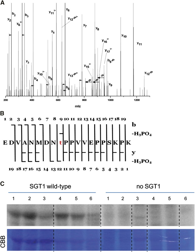
(A) Fragmentation spectrum assigned to the phosphorylated form of the peptide EDVANMDNTPPVVEPPSKPK (Mascot score 126). Loss of H3PO4 is denoted by –P, loss of water is marked by short horizontal arrows, whereas a longer arrow symbolizes pairs of detected signals corresponding to yn and yn-H3PO4. The majority of signals of the tandem mass spectrometry spectra are assigned to the above species. The presence of several yn>11-H3PO4 and b9-H3PO4 ions accompanied by y15, y16, and y17 pinpoints threonine at position 9 as the unequivocal phosphorylation site within the peptide.
(B) Peptide sequence with assigned y, b, y-H2O, b-H2O, y-H3PO4, and b-H3PO4 ions present.
(C) Recombinant maize SGT1 produced in E. coli was incubated in the buffer containing [γ-32P]ATP, and total proteins were extracted from maize seedlings or tassels infected by various U. maydis strains. The samples were fractionated by SDS-PAGE and analyzed with a phosphor imager. Columns 1 to 3, extracts from seedling leaves 6 DPI with U. maydis wild-type SG200 (1), SG200∆see1 (2), or mock-inoculated (3). Columns 4 to 6, extracts from tassel base 9 DPI with U. maydis wild-type SG200 (4), U. maydis-overexpressing Ppit2:see1 (single-copy integration; 5), or U. maydis-overexpressing Ppit2:see1 (multiple-copy integration; 6). Representative data of four independent biological replicates are shown. CBB, Coomassie Brilliant Blue.

