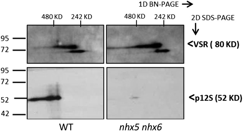
Figure 7.
Separation and Identification of VSR Receptor-Aleurain Cargo Complexes Using 2D BN-PAGE/SDS-PAGE.
Native complexes were separated using 2D BN-PAGE/SDS-PAGE (4 to 16% acrylamide gradient gel) in the first dimension (left to right) followed by SDS (10% acrylamide) in the second dimension (top to bottom) in the wild type (left) and nhx5 nhx6 (right). Forty-five micrograms of total protein from immature seed tissue was loaded on each gel. Immunoblots of anti-VSR1;1 (top) and anti-12S (bottom) are indicated. Signals were quantified from scanned images as described in Methods using four different repetitions of the experiment (one representative set of blots is shown). Wild-type complexes had 3.0 ± 0.9 times more p12S associated with VSR compared with nhx5 nhx6. For each antibody, the same blot exposition time and scan settings were used for the wild type and nhx5 nhx6.

