Figure 8.
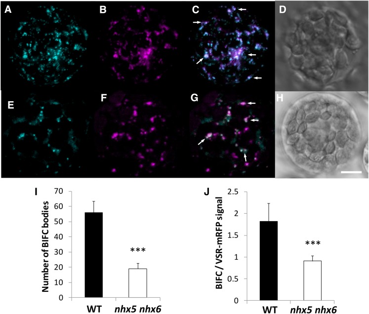
In Vivo Interaction between the VSR Receptor and Its Cargo Protein Aleurain.
Interaction is visualized as BiFC between aleurain fused at the C terminus to the N-terminal half of Venus (Aleu-VYNE) with the C-terminal half of super cyan fluorescent protein (SCYCE) fused to the N terminus of VSR2;1 (SCYCE-VSR2;1) in isolated mesophyll protoplasts. Images are 3D projections of a series of four to five z-stack images and shown in the wild type (A) and nhx5 nhx6 (E). An inactivated VSR1;1-mRFP that properly localizes to VSR-positive compartments was used as a transformation control in the wild type (B) and nhx5 nhx6 (F) (see Methods). (C) shows merged images of (A) and (B) in the wild type, and (G) shows merged images of (E) and (F) in nhx5 nhx6. Differential interference contrast images of the wild type and nhx5 nhx6 are shown in (D) and (H), respectively. (I) and (J) show quantification of the number of BiFC bodies (I) and the BiFC signal intensity relative to the VSR1;1-mRFP control (J). Error bars indicate sd; n = 75. ***P < 0.001, by t test. Bar = 5 μm.

