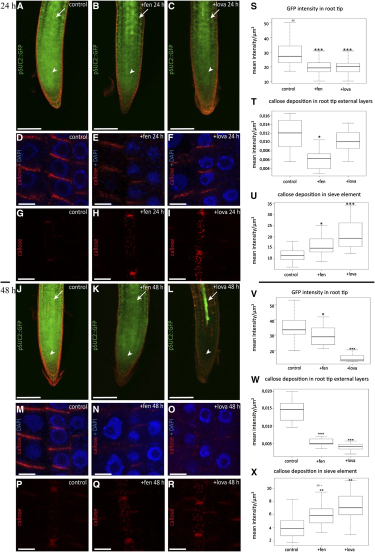
Figure 10.
Altered Sterol Composition Affects Both Symplastic Phloem Unloading and Callose Deposition at PD in Arabidopsis Roots.
(A) to (C), (J) to (L), (S), and (V) GFP symplastic unloading from the phloem to surrounding tissues is reduced in Arabidopsis roots grown on fen ([B] and [K]) or lova ([C] and [L]) for 24 h ([A] to [C]) and 48 h ([J] to [L]). Control seedlings expressing ProSUC2:GFP show diffusion out of the phloem (white arrows) into surrounding tissues including the root meristem (white arrowheads) ([A] and [J]). After fen ([B] and [K]) or lova ([C] and [L]) treatment, GFP transport from companion cells was altered, as indicated by the apparent reduction of the GFP intensity in the root tip and cells surrounding the vasculature. Quantification of fluorescence intensity in the root meristem in control, fen-, and lova-treated plants for 24 and 48 h confirmed that sterol inhibition significantly reduces GFP symplastic unloading from the companion cells to surrounding tissues ([S] and [V], respectively). White arrows indicate the root vasculature where ProSUC2:GFP is expressed, and white arrowheads indicate the GFP unloading zone at the root tip. Bars = 200 μm.
(D) to (I), (M) to (R), (T) to (U), (W), and (X) Callose immunofluorescence (red) in Arabidopsis seedlings treated with fen ([E], [H], [N], and [Q]) or lova ([F], [I], [O], and [R]) for 24 and 48 h. DAPI staining of DNA (blue) was done to highlight the cellular organization of root tissues. Sterol inhibition treatment induced a significant accumulation of callose deposition in sieve elements at both 24 and 48 h, as confirmed by fluorescence quantification ([H] and [U], fen, 24 h; [Q] and [X], fen, 48 h). In external cell layers, callose signal was reduced significantly under 24 h of fen but not lova ([E] and [F], respectively; [T] for fluorescence quantification). However, at 48 h, a strong decrease in callose accumulation was observed in both fen- and lova-treated plants ([N] and [O], respectively; [W] for fluorescence quantification). Bars = 5 μm.
Callose immunolabeling and ProSUC2:GFP experiments were performed at least three independent times using a total of 20 seedlings per condition. Asterisks indicate significant differences (*P < 0.05, **P < 0.01, ***P < 0.001) by Wilcoxon test.

