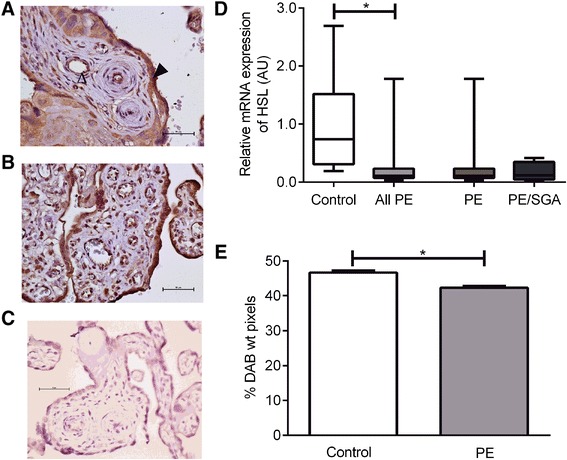
Fig. 2.

HSL. a Immunohistochemisty for the detection of HSL in control placenta, open arrow indicates endothelial cell staining, closed arrow indicates trophoblast staining. b immunohistochemistry in PE placenta. c negative control. d mRNA expression, showing control compared with all PE cases with the result of statistical analysis, as well as the result for PE alone or PE/SGA cases. e Relative protein expression as measured by semi-quantitative immunohistochemistry. *, P < 0.05
