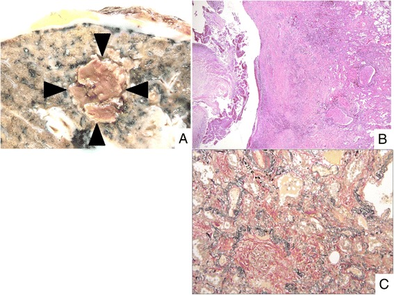
Fig. 2.

Histopathology of organization surrounding the cavity. a Macroscopic findings reveal the fungus ball (arrow head). Pleura with fibrous thickening is also noted. b Organization area can be seen around the fungus ball (H-E staining). c Alveolar spaces are filled with dense collagenous tissue (EVG staining)
