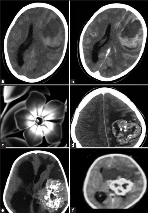
Figure 2.

Supratentorial ependymoma (STE) - Intraparenchymal form: (a) Axial nonenhanced low-dose computed tomography (NECT) and (b) contrast-enhanced computed tomography (CECT). The solid component of the tumor is isodense to gray matter and shows moderate to intense enhancement on CECT. Central nonenhancing areas of necrosis are also seen. The peripheral cystic component shows enhancing margins. Periwinkle sign: (c) Black and White picture of periwinkle flower to which the tumor has been likened to Figure 2d–f. NECT axial images of the intraparenchymal form of STE show the characteristic periwinkle sign due to its lobulated margins (demarcated with a brown line in Figure 2d), central necrosis and centripetal pattern of calcification. Large peripheral cyst is also noted which has been likened to a leaf. This sign is evident with varying degree of calcification as noted in Figures 2d–f
