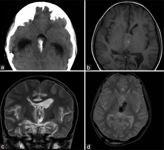
Figure 4.

Supratentorial ependymona-intraventricular form: (a) Axial nonenhanced low-dose computed tomography. The tumor is isodense to gray matter and shows central calcification. Secondary hydrocephalus is also noted. (b) Axial T1-weighted (c) Coronal T2-weighted (d) Axial gradient echo magnetic resonance image of the same case shows tumor is isointense to gray matter on T1 sequence and hyperintense on T2 sequence. Central areas of calcification which are seen as T1 hyperintensity, T2 hypointensity and which are blooming on GE sequences are noted
