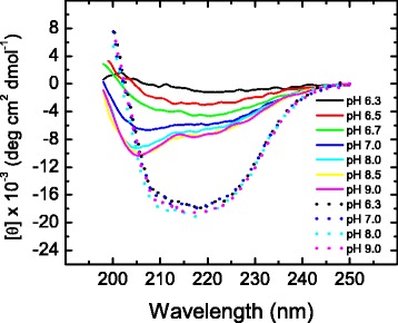
Fig. 2.

Far-UV CD spectra of thermally unfolded yTIM at different pH values. Protein samples were allowed to unfold by continuous heating (2 °C min−1) until the end of the transition (cf. Fig. 1) and then left to stand at 70.0 °C for 10 min before recording their spectra. For comparison, spectra of native yTIM at various pH values are also shown (dotted lines)
