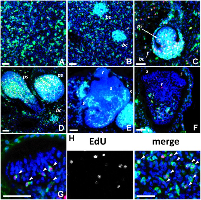
Fig. 7.—
WMISH analysis of ta-TRIM expression during E. multilocularis larval development. In all panels, the ta-TRIM WMISH signal is shown in green, DAPI (all nuclei) in blue, and EdU detection in red (EdU was incorporated during a 5 h, 50 μM pulse, in vitro). Staging follows the system of Leducq and Gabrion (1992). (A) Germinal layer. (B) Early formation of brood capsule buds (bc) from the germinal layer. (C) Early formation of the protoscolex (ps; stage 1). (D) Early formation of the protoscolex (ps; stage 2). (E) Intermediate protoscolex development (stages 3 and 4). r, rostellum; s, sucker primordia. (F) Late protoscolex development (already invaginating, stage 6). r, rostellum (red signal in rostellum comes from auto-fluorescence of the hooks); s, suckers. (G) Detail of the sucker of the protoscolex shown in (F). Arrowheads point at EdU+ ta-TRIM + cells at the base of the developing suckers. (H) Detail of the germinal layer. Arrowheads point at EdU+ ta-TRIM+ cells. Bars, 25 μm.

