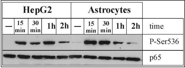
Fig. 8. Transient phosphorylation of p65 on serine 536.

Human astrocytes and hepatoma HepG2 cells were treated with IL-1α (10 ng/ml) and 1 μM DEX for indicated time periods, and cell lysates were prepared. Protein concentrations were determined in the lysates and equal amounts of total cellular protein were analyzed by Western blotting using anti-Phospho-NF-κB p65 (Ser536) or anti-p65 antibodies. Representative results of two experiments are shown.
