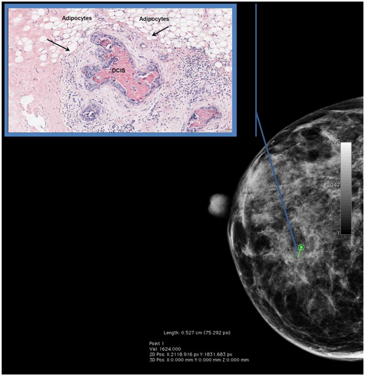
Figure 4.

Peri-lesional mammographic density surrounding a DCIS lesion is increased relative to that of the entire breast (here, peri-lesional fibroglandular volume=85% and overall fibroglandular volume for the entire breast=68%). To compute peri-lesional mammographic density, the radiologist recorded the biopsy location and radius of the biopsy target (noted in green) on a craniocaudal view of the pre-biopsy digital mammogram. Percent dense fibroglandular volume was estimated using single x-ray absorptiometry at a volume twice the size of but excluding the biopsy target, centered at the biopsy site. We suggest that large differences between peri-lesional and total volumetric mammographic density may be associated with DCIS.
Inset: A hematoxylin and eosin stained tissue section of the biopsy target from the same patient shows that a central area of DCIS with necrosis is surrounded by a rim of reactive stroma. In a study of women who were clinically referred for an image-guided breast biopsy at the University of Vermont Breast Cancer Surveillance Consortium site, we are currently investigating whether peri-lesional mammographic density is a useful predictor of underlying pathology.
