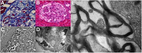
Fig. 2.

Cytoplasmic vacuolation observed in a podocyte by light microscopy with a Masson’s trichrome and b Haematoxylin and eosin staining. c Myelin-like structures in a podocyte, with concentric lamellated ultra-structural appearance. (Electron Microscopy). d Focal areas of podocyte effacement; amorphous myelin-like structures are visible in glomerular parietal epithelial cells and in endothelial cells (Electron Microscopy). e Myelin-like structures parallel with zebra-like body appearance (Electron Microscopy)
