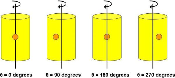
Figure 2.
Diagram showing the object geometry for our example case study. The object is a mouse-sized cylinder with an interior 5 mm diameter spherical tumor offset 5 mm from the center of rotation of the object. The orientation angles correspond to the angles in Figures 3(a)-(f). The reader is viewing the images as if she/he were the detector (i.e. the tumor is closest to the detector at θ=0 degrees).

