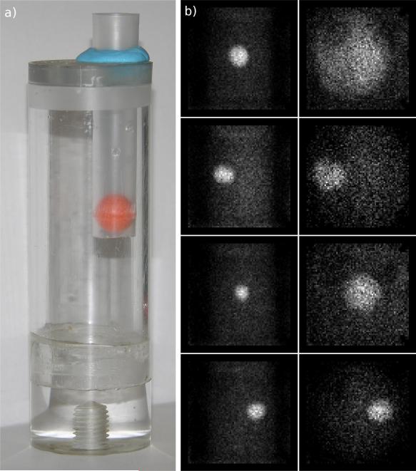
Figure 5.
a) Phantom used for imaging study. The large cylinder holds the background source while the orange sphere holds the lesion source. The orange sphere is a plastic, hollow ball. b) (left) Projection images from fixed geometry data set (right) Analogous projection images from adaptive data set.

