
Fig. 1.
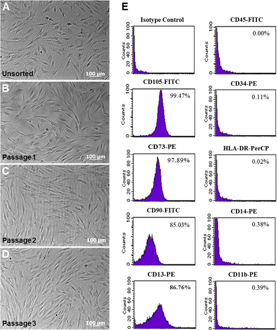
Cell culture and flow cytometry analysis of isolated dental pulp stem cells (DPSCs). a Representative phase-contrast micrographs shows unsorted-cells derived from human dental pulp tissue after 14 days of cell culture. b–d CD105+ magnetically-sorted DPSCs cultured in α-MEM without osteogenic induction. Morphologically, cells appear as typical fibroblastic and spindle shape during 3 passages. Original magnification 10x, scale bar =100 μm. e Flow cytometric analysis presented as histograms that show cell fluorescence intensity on the horizontal axis and cell frequency distribution on the vertical axis. Percentage results show positive expression to immunophenotype associated with mesenchymal stem cell (MSC) lineage as well as a lack of expression for hematopoietic markers