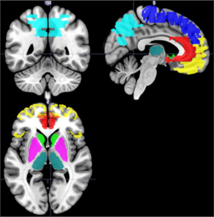
Figure 1.
Brain image showing the regions of interests used for analysis. Regions of interest have been labelled using coloured fills. Yellow: orbito-frontal cortex; dark blue: medial frontal gyrus; red: anterior cingulate gyrus; light green: caudate; cyan: lenticular nucleus; dark green: thalamus; light blue: superior parietal lobiule and precuneus.

