
Figure 1.
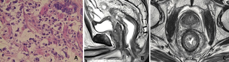
Pathology and MRI features in case 1. (A) Rectal biopsy shows scattered signet-ring cells (Hematoxylin and eosin staining, ×20). (B) T2-weighted sagittal plane MR image shows a diffuse wall thickening of the rectum over 15 cm. (C) T2-weighted axial plane shows a circumferential thickening over 11 mm with a concentric ring pattern.