
Figure 2.
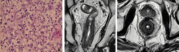
Pathology and MRI features in case 2. (A) Scattered signet-ring cells (Hematoxylin and eosin staining, ×20). (B) Pelvic MRI shows on T2-weighted coronal plane a diffuse wall thickening of the entire rectum over a length of 18 cm, accounting for its markedly rigid tube aspect. (C) On T2-weighted axial plane, a circumferential wall thickening over 13 mm with infiltration of the mesorectum and a concentric ring pattern.