
Figure 3.
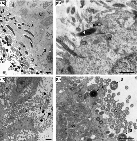
Transmission electron microscopy showing spermatozoal attachment in the oviduct of Chinese soft-shelled turtle. (A) Many spermatozoa (S) were embedded among cilia (Ci) and inserted into the apical hollowness of the ciliated cell (CC). (B) A spermatozoon inserted its head (H) into the hollowness which was surrounded by no lysosome in ciliated cell. (C) Spermatozoa were present at the opening of gland conduit. (D) Cross section of the spermatozoa head in the hollowness of ciliated cell and the spermatozoa midpiece among cilia. basal body (B), secretory cell (SC), mitochondrion (Mi). Scale bar = 2 μm (A, C) and 1 μm (B, D).