
Figure 5.
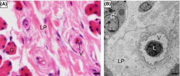
Few immune cells occasionally appeared inside the blood vessel of the lamina propria. (A) The blood vessel in the mucosa of infundibulum, H-E stain. (B) Lymphocyte inside the blood vessel of the lamina propria (LP) in the uterine, TEM. oviduct gland (G), lamina propria (LP), lymphocyte (L), blood vessel (V). Scale bar = 10 μm (A) and 2 μm (B).