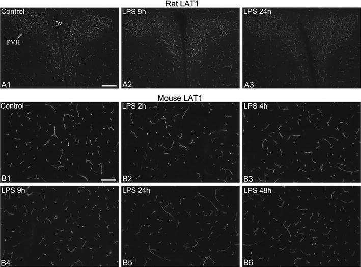
Fig. 6.

LAT1 immunofluorescence in brain vessels of the rat and mouse hypothalamus. A LAT1 immunofluorescence in the rat hypothalamus at the level of the paraventricular nucleus (PVH). The intensity of labeling tended to be lower at 24 h after LPS administration. Scale bar 200 µm. B LAT1 immunofluorescence in hypothalamic vessels of mice; labeling intensity is modestly reduced at 24 and 48 h after LPS. Scale bar 100 µm
