
Fig. 7.
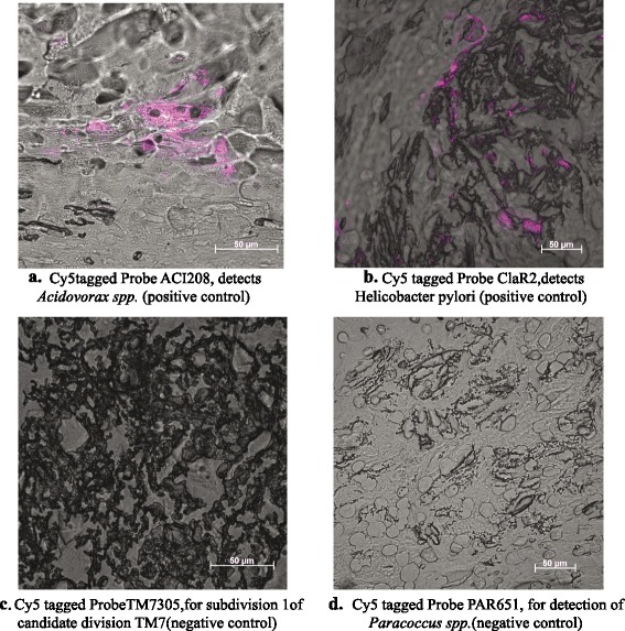
FISH images of Acidovorax spp. and H. pylori in atherosclerotic plaque samples. Representative results of plaque samples after applying the Acidovorax spp.-specific probe (a), H. pylori-specific probe (b), as well as two negative control probes, i.e., Candidate division TM7 (c), and Paracoccus spp. (d), on atherosclerotic plaque material from several different patients. We observed morphogically distinct clusters of Acidovorax spp. and H. pylori cells surrounded by atherosclerotic tissue material