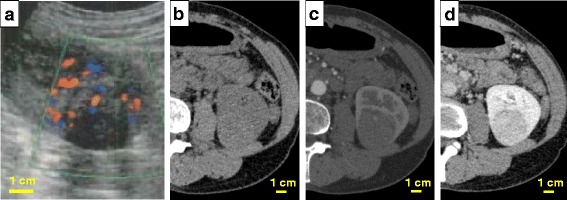
Fig. 1.

Clinical images of the left renal tumor. A mass with clear margin and mostly endophylic growth was detected in the lower and posterior part of the left kidney. a The tumor presented with a non-cystic mass of a heterogeneous nature in ultrasound. Color Doppler imaging showed hypervascularity in the tumor. b, c, d) Dynamic computed tomography of the left renal tumor. b Image before administration of the contrast medium. c Early phase of enhanced computed tomography. A blood vessel was visualized in the tumor. d The tumor showed earlier washout of enhancement than the adjacent normal renal parenchyma
