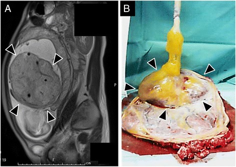
Fig. 1.

Clinical findings of cord hemangioma. a Fetal magnetic resonance imaging (MRI) performed at 27th week of gestation. The solid mass lesion was identified close to but not attaching to the placental disc. Note the umbilical vessels are identified within the mass. b Macroscopic appearance of umbilical cord and placental disc before formalin fixation. The large and solid mass is seen at the placental end of the umbilical cord (Arrow heads)
