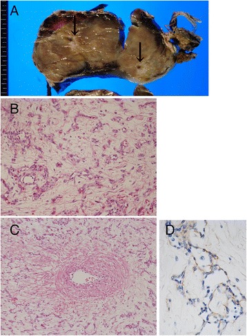
Fig. 2.

Pathological findings of cord hemangioma. a Cut surface of cord hemangioma, showing a tan-colored and solid mass containing umbilical vessels (arrows). b Representative histology of the cord hemangioma. The tumor consists of small, arborizing and thin-walled vessels (Hematoxylin and eosin staining). c Original umbilical artery (left side) passes through the tumor. The tumorous vessels never invade to the original umbilical vessels. d Endothelial cells of the tumor are immunohistochemically positive for AFP
