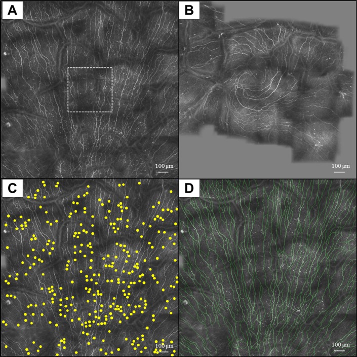
Figure 1.
Wide-field composite IVCM images of the subbasal layer in the central cornea (A, B). The composite image is significantly larger compared with a standard image (dashed box in [A]). Use of the Cell Counter plug-in of ImageJ (C) and the NeuronJ (D) for measuring the DC and nerve densities, respectively.

