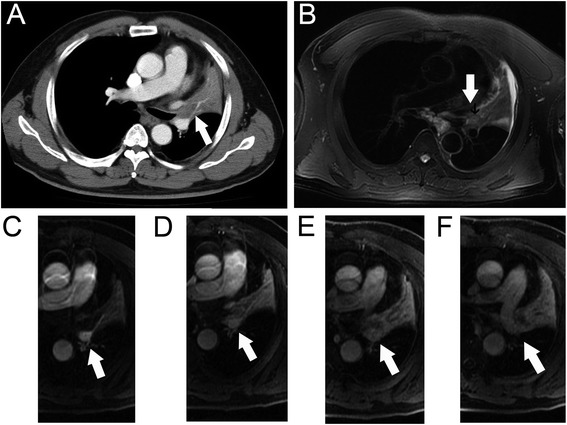
Fig. 1.

Images of 64-year-old man with diagnosed squamous cell carcinoma of the left upper lung. The extent of primary tumor in the left hilum (arrow) was not accurately distinguished from the secondary changes on axial MDCT (a). Tumor (arrow) was appeared as slightly hypointense compared the signal of secondary changes on axial T2-weighted MRI (b). Tumor (arrow) was showed as hypointense while that of secondary changes were hyperintense in early phase of dynamic contrast-enhanced MRI (c and d), in delay phase both of them were appeared as heterogeneously hyperintanse (e and f)
