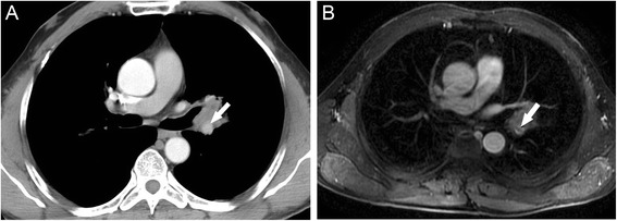
Fig. 4.

Images of 50-year-old woman with diagnosed adenocarcinoma of the left lung. Left pulmonary artery was visualized being encased with tumor tissue at less than 180° (arrow) which indicated no involvement of the vessel on transverse MDCT (a). Transverse contrast-enhanced MR image showed filling defect within the lumen of left pulmonary artery (arrow), caused the concern of tumor extending into the artery (b). The invasion was then confirmed with pathology (tumor was staged T4)
