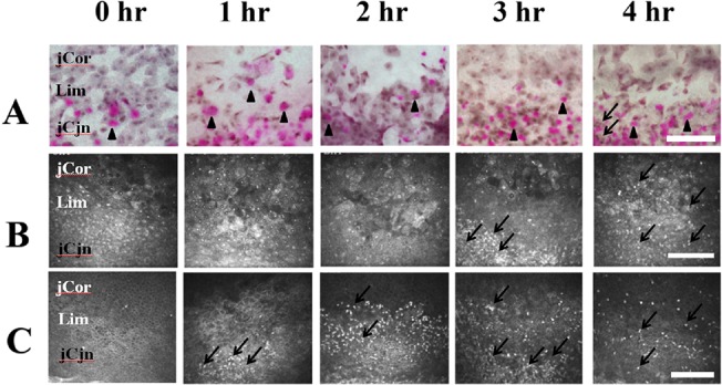
Fig 1. Impression cytology and in vivo confocal microscopy of the limbal epithelium.

(A) Impression cytological findings. At post exposure 1 to 4 hours, the morphology of the superficial epithelial cells and goblet cells (black arrow heads, the purple dots indicate goblet cells containing mucin which were stained by PAS) attached to the filter papers were similar. At post exposure 4 hours, some inflammatory cells (black arrows) can be found at the juxta-limbal conjunctival site. (B) In vivo confocal microscopic findings of the surface epithelial layer of the limbus. there was no significant change of morphology of superficial cells from post exposure 1 to 4 hours. An aggregation of inflammatory cells (black arrows) can be found atpost exposure 3 hours to 4 hours. (C) In vivo confocal microscopic findings of the basal epithelial layer of the limbus. There was no significant change of basal cell morphology from post exposure 1 to 4 hours. Aggregation of inflammatory cells (black arrows) can be found at post exposure 1 and 2 hours. The inflammatory cells migrated to the juxta-limbal corneal and juxta-limbal conjuncival areas at post-exposure 3 and 4 hours.jCor: juxtal-limbal cornea. Lim: limbus. jCjn: juxta-limbal conjunctiva. hr: Hours after exposure. Bar: 200 um.
