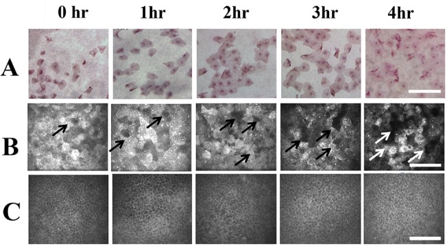
Fig 4. Impression cytology and in vivo confocal microscopy of the central cornea epithelium.

(A) Impression cytological findings. The longer time the cornea exposure, the more surface cells attached to the filtering paper were found. However, the morphology of the detached cells remained similar from post-exposure 1 hour to 4 hours. (B). In vivo confocal microscopic findings of the surface epithelial layer of the central cornea. From 1 hour to 4 hours after exposure, the time dependent increase of cellular disappearance (White and black arrows) was found, which indicated the sloughing of the cell. (C) In vivo confocal microscopic findings of the basal epithelial layer of the central cornea. There was changes of cellular morphology or density from post-exposure 1 to 4 hours. During the observational period, there was no inflammatory cells detected at the superficial or the basal epithelial layers. hr: Hours after exposure. Bar: 200 um.
