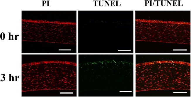
Fig 8. TUNEL staining for the detection of apoptotic cells in the central cornea.

At post exposure 3 hours, the TUNEL stain positive cells can be detected in the corneal epithelium and superficial keratocytes. Green: TUNEL, which stains apoptotic cells. Red: PI, which stains nucleus. Scale bar: 100 um. hr: Hours after exposure.
