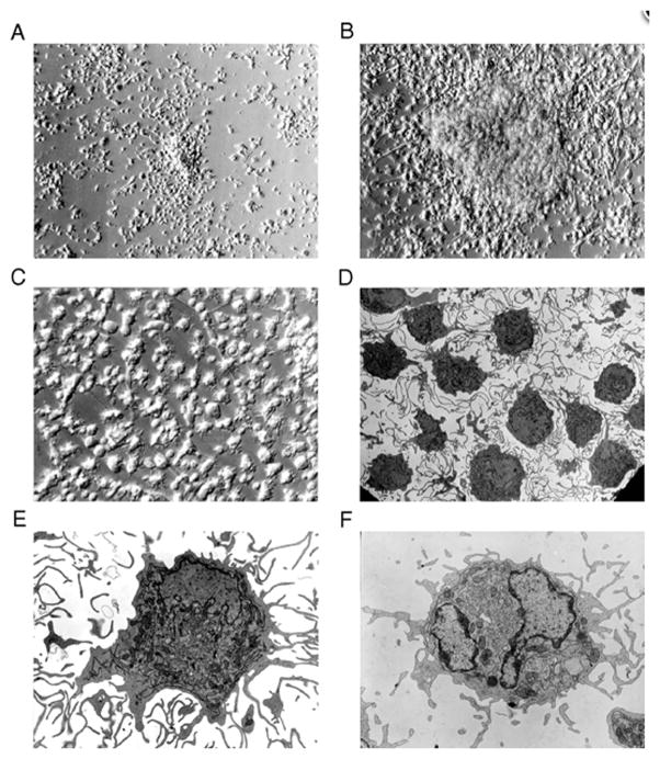
Figure 7.32.1.
Generation and morphology of CD83+ dendritic cells. Monocytes cultured with GM-CSF/IL-4/TNF-α develop a dendritic cell morphology (A–C). Monocytes (CD14+) were isolated and cultured with cytokines for 2 (A) or 5 (B, C) days and examined by phase-contrast microscopy (A–B, 200× magnification; C, 400×). Representative transmission electron micrographs of (D–E) monocyte-derived CD83+ cells isolated by cell sorting from 7-day GM-CSF/IL-4/TNF-α cultures and (F) blood CD83+ dendritic cells (D, 1855× magnification; E-F, 5800×).
This figure, originally published in Zhou and Tedder (1996), is reprinted by permission of Proceedings of the National Academy of Sciences.
Representative phenotype of immature and mature dendritic cells. Phenotype and scatter profiles of monocyte-derived immature DCs (A). Phenotype and scatter profile of monocyte-derived mature DCs (B). Data is depicted as a 2-color analysis using PerCP-labeled class II versus PE-labeled antibodies as indicated in the figure. Peridinin Chlorophyll Protein Complex, PerCP; Phycoerythrin, PE.

