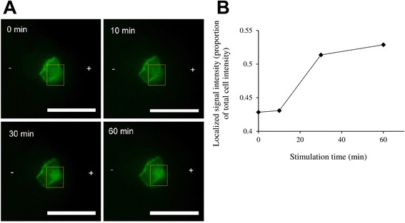
Fig. 3.

Polarization of PH-Akt-GFP in HaCaT cells under DC stimulation (150mV/mm). a Over time, localization of PH-Akt-GFP to the anodal side (+) of the cell was observed. b The localized signal intensity at the anodal end of each cell (yellow dotted box) was quantitated as a proportion of the total signal intensity of the cell. Scale bar = 50 μm
