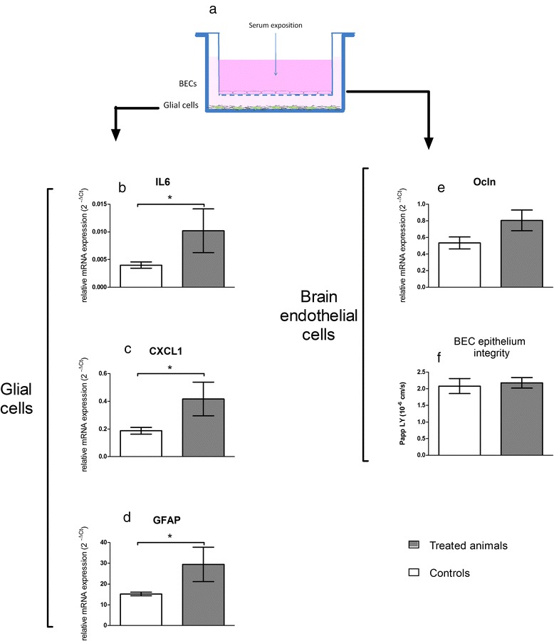
Fig. 11.

Impact of exposure to sera from treated animals on the in vitro BBB model. (a) is a schematic view of the model architecture. BECs were grown on a semipermeable membrane whereas glial cells lay on the well bottom. mRNA expressions were quantified by RT-qPCR after 24 h exposure to sera from control and treated rats. For glial cells, expressionsof cytokines and chemokines (IL6 (b), CXCL1 (c)) and of GFAP (d) are presented. For BECs, tight junction protein occludin (Ocln (e)) is reported and integrity of the BECs monolayer was checked in terms of apparent permeability to Lucifer yellow (f). Sera diluted in culture medium were applied to the BECs compartment for 24 h. Each data point represents the mean ± SEM of n = 4 samples, each sample representing the mRNA pool from 6 wells. RT-qPCR was performed in duplicate for each single sample. Statistical comparison was performed by one tailed Mann Whitney test, * P < 0.05 for IL-6, CXCL1 and GFAP mRNA expressions. Statistical comparison was performed by two tailed Mann Whitney test for Ocln mRNA expression (P = 0,057)
