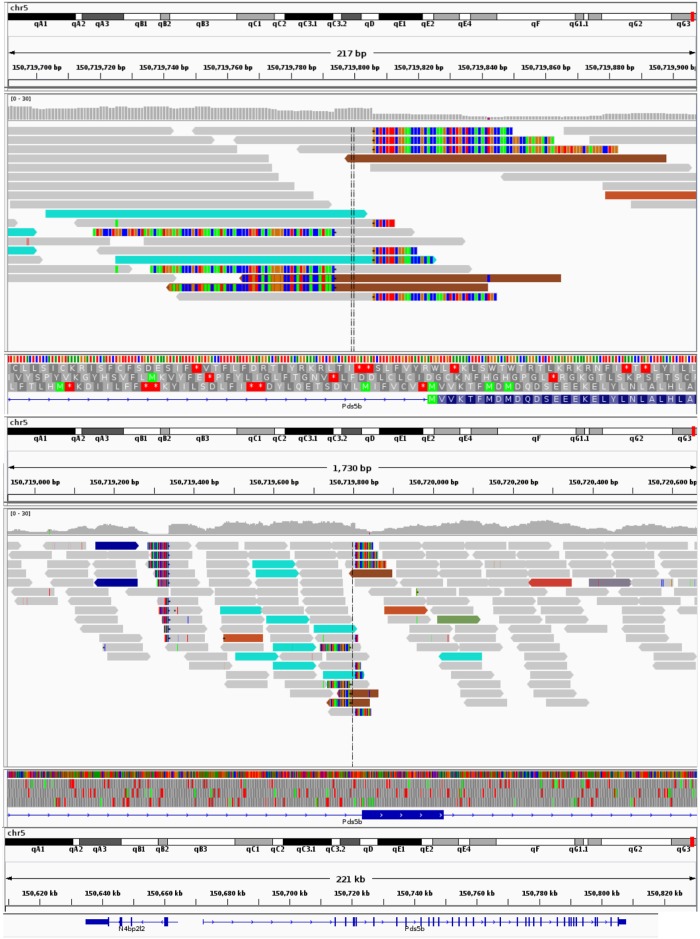
Fig 3. Read alignments surrounding transgene integration.
The screenshots from the IGV genome viewer show the integration site of the transgene on chromosome 5. A) displays the site and its surroundings. Grey reads are concordantly mapped reads. The colored reads indicate that the other half of the fragment maps discordantly. The teal colored reads are those that have mates on the Her2 transgene. Multi-colored segments at the ends of reads highlight soft-clipped portions of reads. There are three clusters of soft-clipped reads. Two around the integration site, and another block upstream that is unrelated to the integration. B) shows the integration in more detail. The proximity to exon 3 of the gene can be seen at the bottom. The right block of soft-clipped reads contains sequence that corresponds to just upstream of the transgene coordinate 4025 (the insert). The left block’s sequence derives from transgene coordinate 2972 and marks the return to chromosome 5 at the end of the concatemer.

