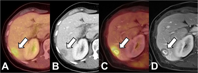
Fig 2. Patient with breast cancer.

Both PET/CT (A,B) as well as PET/MRI (C; D; VIBE, portal venous phase) show a lesion with elevated FDG-uptake and ill-defined lesion borders as well as central contrast enhancement as signs of malignancy. Based on these findings the lesion was correctly identified as metastasis in both modalities.
