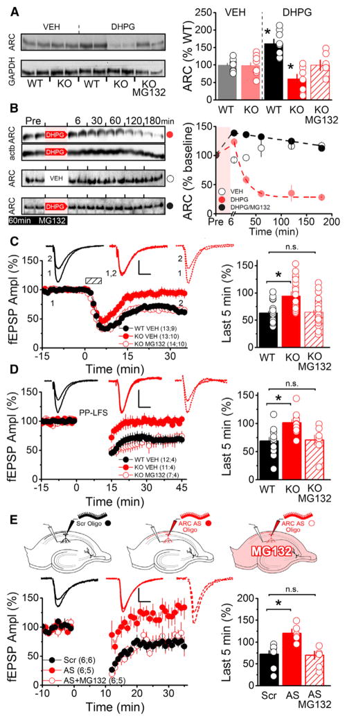
Figure 1. Proteasome Inhibition Rescues mGluR-Triggered Translation of ARC and mGluR-LTD.
(A) Hippocampal slices from Sam68 KO or WT mice were treated with vehicle (VEH) or 50 μM (R,S)-3,5-DHPG for 5 min. (S)-MG132 (5 μM) was added for 1 hr before and during DHPG treatment. (Right) Densitometric quantification of western blots normalized to GapDH. Each dot represents a separate experiment consisting of pooled lysate from three slices. The average for each condition was generated from eight separate experiments using slices from four KO and WT mice. An asterisk denotes a significant difference from WT (VEH). ([VEH] WT, 100.0 ± 4.2; KO, 99.0 ± 6.8; [DHPG] WT, 161.6 ± 12.7; KO, 60.5 ± 13.1; KO [MG132], 99.2 ± 14.9).
(B) Western blots (left) and scatter plot (right) showing time courses of protein levels after three different treatments. (Left) Rat hippocampal slices were treated with 100 μM DHPG for 6 min (top, ARC [red circles] and actb [not plotted]); vehicle (middle, ARC [open circles]) or 5 μM MG132 for 1 hr before and during DHPG treatment (bottom, ARC [black circles]). Each time point represents pooled lysate from three slices. (Right) Time course plot showing DHPG application (shaded red area) results in sustained depletion in ARC protein levels that is blocked by proteasome inhibition. Each dot represents average ARC levels at indicated time points (n = 4 rats for DHPG, n = 2 rats for VEH and MG132/DHPG).
(C) Extracellular field recordings (fEPSP) in acute hippocampal slices show Sam68 KO mice (red filled circles) lack mGluR-LTD at Schaffer collateral synapses induced by 50 μM DHPG for 5 min. MG132 (5 μM) for 1 hr before and during DHPG treatment rescued LTD (red open circles). Dashed box indicates time of DHPG application. Scale bars indicate 10 ms and 0.25 mV. (Right) Average fEPSP slope during the last 5 min of recording (WT, 59.4 ± 3.9; KO, 91.9 ± 8.1; KO MG132, 59.8 ± 3.9).
(D) Sam68 KO mice lack mGluR-LTD induced by PP-LFS for 15 min (2 pulses at 50 ms ISI, 1Hz). MG132 (5 μM) for 1 hr before and during PP-LFS rescued LTD (WT, 67.7 ± 7.3; KO, 99.8 ± 6.5; KO MG132, 70.4 ± 8.1).
(E) Whole-cell recordings in voltage clamp (−60 mV) in acute hippocampal slices (mouse) with an antisense oligonucleotide against ARC (AS, 150 μM, filled red circles) or a scrambled oligo (Scr, 150 μM, filled black circles) loaded into the patch pipette. Proteasomal inhibition rescues mGluR-LTD blocked by acute knockdown of ARC synthesis by antisense oligonucleotides (AS+MG132, red open circles) (Scr, 72.2 ± 8.2; AS, 120.3 ± 9.5; AS+MG132, 70.0 ± 9.1).
Summary data consist of mean ± SEM.

