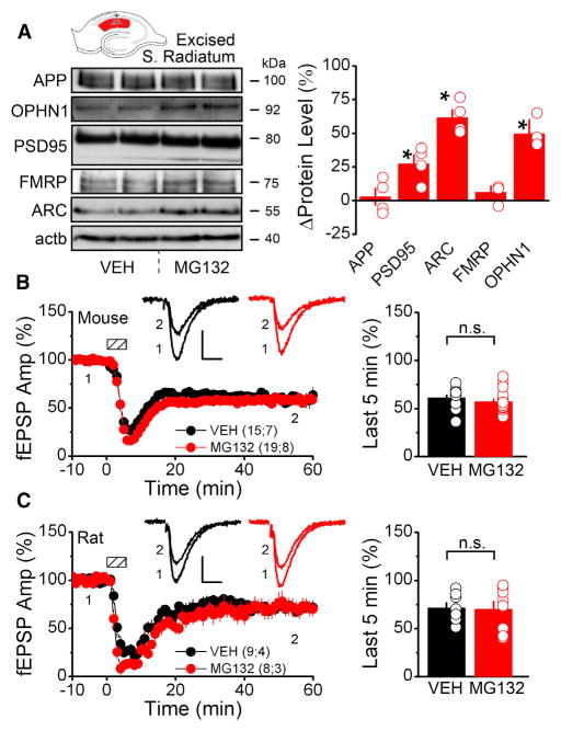
Figure 2. Proteasomal Inhibition Increases Dendritic PRP Levels with No Change in mGluR-LTD Magnitude.
(A) Western blots of PRPs from rat microdissected S. Radiatum incubated in 5 μM MG132 for 1 hr or vehicle (APP, amyloid precursor protein; OPHN1, oligophrenin 1; PSD95, post-synaptic density 95; FMRP, fragile X mental retardation protein; ARC, activity-regulated cytoskeletal-associated protein; actb, β-actin). (Right) Densitometric quantification shows brief proteasome inhibition significantly increases dendritic levels of PSD95, ARC, and OPHN1 compared with vehicle condition (APP, 3.1 ± 6.5; PSD95, 27.4 ± 7.0; ARC, 61.5 ± 5.9; FMRP, 7.0 ± 4.7; OPHN1, 49.7 ± 10.4). The asterisk denotes significantly different than vehicle.
(B and C) MG132 does not affect mGluR-LTD at Schaffer collateral synapses in acute hippocampal slices. Dashed boxes show time of DHPG application (mouse, 50 μM, 5 min; rat, 100 μM, 6 min). MG132 (5 μM) for 1 hr before and during DHPG treatment does not change mGluR-LTD magnitude in mice (B) or rats (C). Scale bars indicate 10 ms and 0.25 mV. (Right) Average fEPSP slope during the last 5 min of the recording ([mouse] VEH, 61.3 ± 2.6; MG132, 57.2 ± 2.8; [rat] VEH, 71.6 ± 4.7; MG132, 70.2 ± 8.1).
Summary data consist of mean ± SEM.

