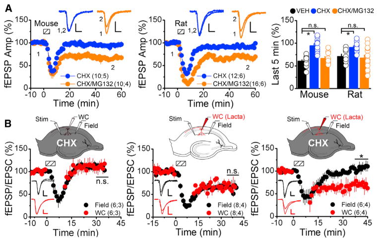
Figure 3. mGluR-LTD Persists in the Absence of Translation if the Proteasome Is Inhibited.
(A) Field recordings from acute hippocampal slices (mouse and rat) treated with either 60 μM CHX (blue) or 60 μM CHX and 5 μM MG132 (CHX/MG, orange) for 1 hr. Dashed boxes show time of DHPG application (mouse, 50 μM, 5 min; rat, 100 μM, 6 min). Scale bars indicate 10 ms and 0.25 mV. (Right) Average fEPSP slope during the last 5 min of recording indicates inhibiting the proteasome rescues mGluR-LTD blocked by translational inhibition ([mouse] VEH, 61.3 ± 2.6; CHX, 94.7 ± 4.7; CHX+MG132, 66.9 ± 3.9; [rat] VEH, 71.6 ± 5.2; CHX, 91.2 ± 3.3; CHX+MG132, 67.7 ± 6.0).
(B) Concurrent field (black) and whole-cell recordings (red) of mGluR-LTD (50 μM DHPG, 5 min) from mouse slices. (Left) CHX (60 μM) included in the perfusate blocked LTD (WC, 109 ± 12; field, 110 ± 6). (Middle) Lactacystin (1 μM) included in the recording pipette, with vehicle in the perfusate resulted in no significant difference in the amount of LTD between the field and whole-cell recordings (WC, 68.5 ± 7.7; field 67.7 ± 6.9). (Right) Lactacystin (1 μM) in the recording pipette and 60 μM CHX in bath blocked LTD in the field recordings, but not in the whole-cell recordings (WC, 68.7 ± 11.5; field, 108.8 ± 10.3). Scale bars indicate 0.25 mV/50 pA and 10 ms.
Summary data consist of mean ± SEM.

