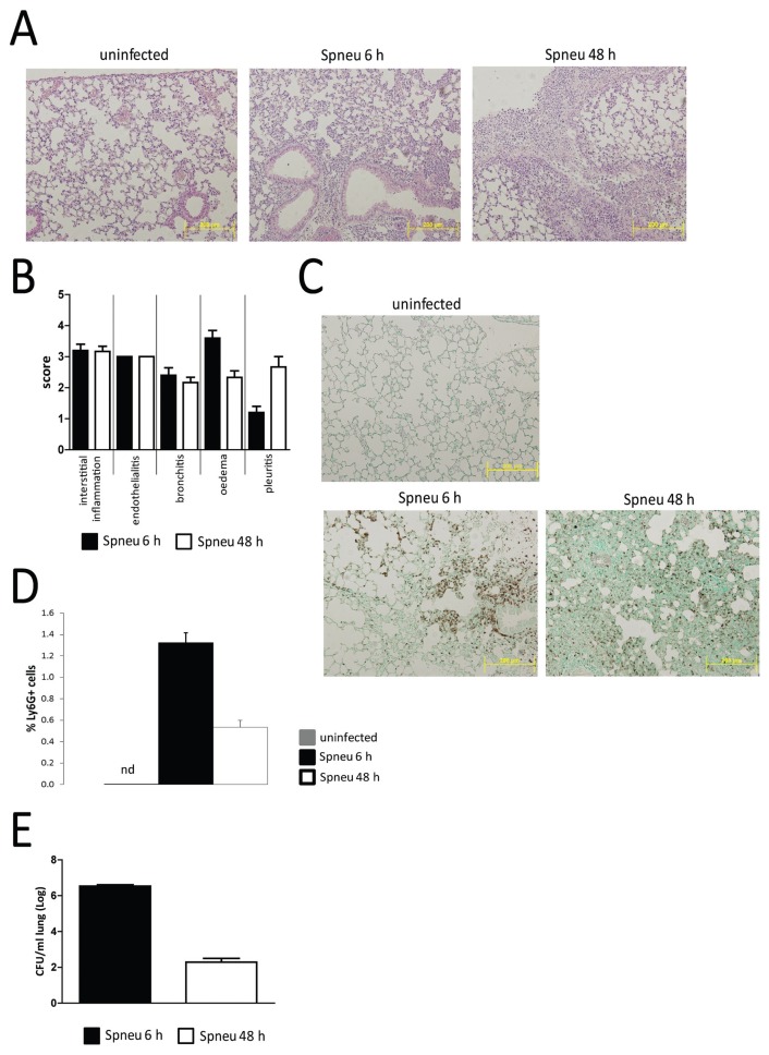
Figure 1.
Immunohistochemical analysis of the early and late host pulmonary response to pneumococcal infection. C57BL/6 female mice were each intranasally inoculated with 2 × 10⁷ CFUs S. pneumoniae D39. Mice were killed at 6 or 48 h (n = 4 each) and lungs were used for histological examination. (A) HE-stained representative lung sections of noninfected (C57BL/6; n = 3), 6-h postinfection (Spneu 6 h) and 48-h postinfection (Spneu 48 h) mice. (B) Histological scoring, determined as described in Materials and Methods, showed inflammatory pathology was largely similar between 6 h and 48 h. (C, D) Granulocyte abundance was higher at 6 h postinfection than at 48 h postinfection, evidenced by higher anti-Ly6G+ cell counts. (E) Bacterial burden measured as CFUs/mL was higher at 6 h postinfection than at 48 h postinfection. Data represent means ± SEM.

