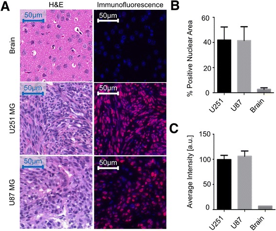
Fig. 1.

PARP1 immunofluorescence staining of brain, U251 MG xenografts, and U87 MG xenografts. a Representative images showing H&E staining (left) and PARP1 immunofluorescence staining (right) of each tissue type. b Percent of nuclei positive for PARP1 for each tissue type. c Average intensity of nuclei in each cell type
