Fig. 3.
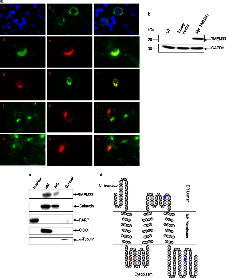
TMEM33 is an ER transmembrane resident protein. a Immunofluorescence analysis of ER localization of TMEM33 in COS-1 transfectants. After 48 h of transfection with Myc-TMEM33 cDNA expression vector, COS-1 transfectants were fixed and immunostained as described in “Materials and methods” section. For ER-tracker or Mito-tracker staining, transfected cells were first incubated with these dyes. Right subpanels: a DAPI; d anti- Calnexin antibody; g ER-tracker; j Mito-tracker; m, WGA. Middle subpanels (b, e, h, k, n): anti-Myc antibody. Left panels (c, f, i, l, o): merge of corresponding right and middle panels. b Expression of Myc-TMEM33 protein in transiently transfected COS-1 cells was confirmed by immunoblotting with anti-Myc antibody. The blot was reprobed with anti-GAPDH antibody. UT untransfected. c The subcellular localization of endogenous TMEM33. MCF-7 cells were homogenized and fractionated into nuclear, heavy membrane (HM), microsomal (MS), and cytosolic fractions, followed by sequentially immunoblotting of the cell fractions using anti-TMEM33, anti-Calnexin (ER membrane), anti-PARP (nucleus), anti-COX4 (mitochondria), and anti-tubulin (cytosol) antibodies. WCE, whole cell extract. d The ER membrane structure of TMEM33. TOPO2, transmembrane protein display software, was used to display 2D topology of TMEM33. ER lumen, N-terminus 1–31 aa; 122–155 aa; ER membrane, 32–52 aa; 101–121 aa; 156–176 aa; Cytoplasm, 53–100 aa; 177–247 aa—C terminus; Potential phosphorylation site (T65) (red) and two ubiquitination sites (K148 and K221) (blue) are shown

