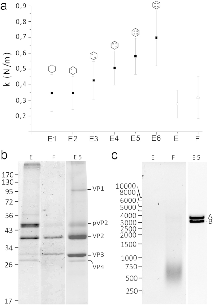
Figure 2. Comparison of the mechanical rigidity of E1-E6 IBDV capsids with empty and full IBDV VLP.
(a) Mean rigidity values of E1-E6 IBDV capsids, as well as empty (E) and full (F) IBDV-derived T = 13 virus-like particles. (b) Coomassie blue-stained SDS-PAGE gels of empty (E) and full (F) VLP, and E5 capsids. Molecular size markers (× 10−3 Da) are shown at left; bands corresponding to proteins VP1, pVP2, VP2, VP3 and VP4 are indicated. (c) Agarose gel electrophoresis of nucleic acids contained in empty (E) and full (F) VLP, and in E5 particles. Cell ssRNA is seen as a diffuse band (F), and bands of dsRNA A and B segments are indicated. Left, molecular weight markers (bp). Full-length gels are presented in Supplementary Fig. 3.

